
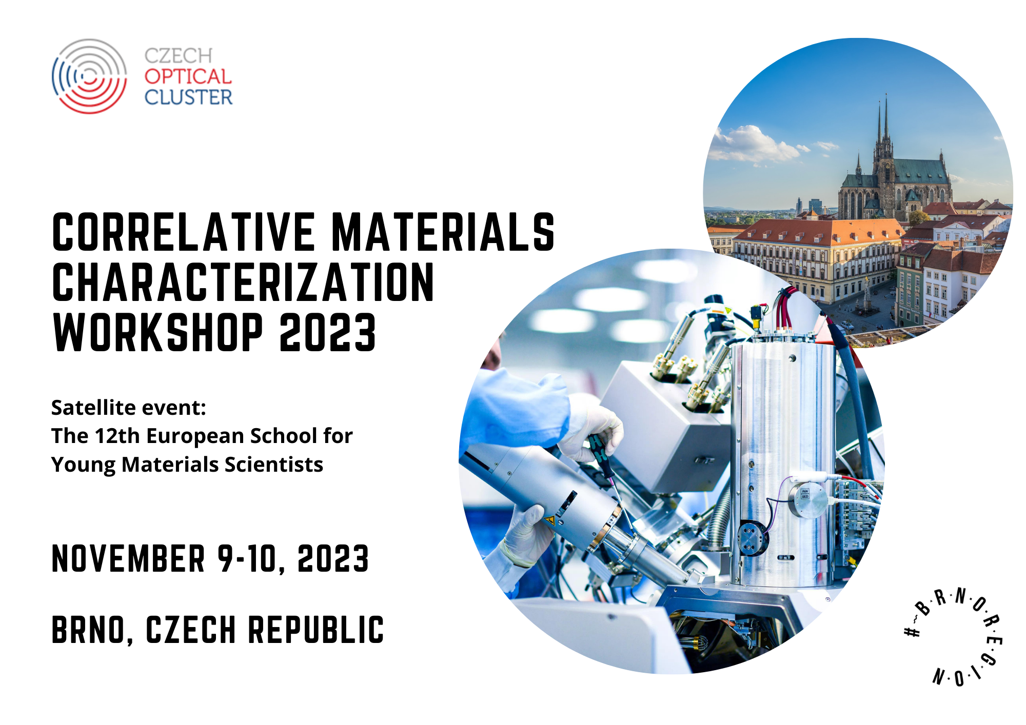
Correlative Materials Characterization Workshop 2023 – Brno, Czech Republic
The workshop will be dedicated to the rapidly growing field of Correlative Materials Characterization and Correlative Imaging of living and non-living systems. The workshop’s scope includes all relevant techniques used for correlative materials and biomaterial characterization. The focus will be on Light Microscopy, Electron Microscopy, X-ray Microscopy, Atomic Force Microscopy and others.
The event follows its successful previous edition held in Dresden and aims to bring together communities from Materials, Life and Computer Science to discuss and explore topics of common interests of Correlative Characterisation and related phenomena.
Venue: Brno Observatory and Planetarium
Participation at the event is free of charge, but registration is compulsory.
Program 2023
CMC Workshop Program
CMC-Workshop-Program.pdfThursday - 9.11.2023:
12:30 - 12:40 (10 min) - Welcome - Tomáš Šikola, Jan Neuman, Petr Přikryl - Organizers
12:40 - 12:50 (10 min) - Regional Microscopy Innovation Ecosystem - Petr Střelec, Radka Martínková, Markéta Shánělová - Brnoregion Microscopy Platform
12:50 - 13:10 (20 min) - Correlative Microscopy: from Data to Information - Dirk van der Wal - TESCAN Group
Correlative On-Surface analysis - chair Jan Neuman
13:10 - 13:30 (20 min) - The eukaryotic proteome as a driver for multi-correlative method development - Adrian Nievergelt - Max Planck Institute, Dresden
13:30 - 13:50 (20 min) - Exploring the sub-picoscale in forces and distances - Franz Giessibl - University of Regensburg
13:50 - 14:00 (10 min) - Advanced Nanoscale Chemical Analysis using Photothermal AFM-IR & Correlated Mapping of Mechanical Properties - Dušan Novotný - Burker / Měřící Technika Morava
14:00 - 14:20 (20 min) - 3D multiphase SEM-AFM correlative microscopy - Bohuslav Rezek - CTU
14:20 - 14:40 (20 min) - Light, tips, and molecules: SPM on the path to direct nano-optical measurements - Martin Švec - FZU CAS
14:40 - 15:10 (30 min) – Break
Correlative Tomography and cryo-EM – chair Pavel Tomancak
15:10 - 15:30 (20 min) - Enterovirus cell entry and genome delivery - Pavel Plevka - CEITEC MU
15:30 - 15:50 (20 min) - Tomographic Imaging Capabilities with hard X-Rays at BAMline (Bessy II) - Henning Markötter - BAM Berlin
15:50 - 16:00 (10 min) - High-resolution 3D imaging of materials and structures - Ehrenfried Zchech - deepXscan
16:00 - 16:20 (20 min) - Correlating Structure and Dynamics to Understand Nuclear Self-Assembly - Alexander von Appen - MPI-CBG
16:20 - 16:30 (10 min) - X-Ray Microscopy as a correlative imaging technique - Mohsen Khoshkhoo - ZEISS
16:30 - 16:50 (20 min) - AI in microscopy: Coping with multiple scales and multiple modalities - Michal Kozubek - Masaryk University
16:50 - 17:10 (20 min) - Unlocking Insights: X-ray Micro Computed Tomography as a Correlative Imaging Technique - Jakub Lázňovský - CEITEC BUT
17:10 - 18:00 (50 min) - Poster Session
19:00 - 22:00 - Workshop Dinner
Friday 10.11.2023:
Correlative in-situ analysis – chair Ehrenfried Zchech
9:00 - 9:20 (20 min) - Monitoring dynamic reactions inside the scanning electron microscope - Miroslav Kolíbal - CEITEC BUT
9:20 - 9:30 (10 min) - In-Situ Correlative Microscopy by AFM-in-SEM - Jan Neuman - NenoVision
9:30 - 9:50 (20 min) - Cellulose nanocrystals: from self-assembly to electronic, photonic and energy harvesting devices inspired by nature - Luís Pereira - CENIMAT
9:50 - 10:10 (10 min) - Thermo Fisher Scientific
10:10 - 10:30 (20 min) - Insulating and Metallic Nanostructured Vanadium Dioxide Probed by Electron Energy-Loss Spectroscopy - Andrea Konečná - CEITEC BUT
10:30 - 10:40 (10 min) - Empowering innovation in science with TESCAN solutions - Michal Klášterecký - TESCAN
10:40 - 11:10 (30 min) – Break
Correlative Multimodal and multiscale analysis – chair Tomas Sikola
11:10 - 11:30 (20 min) - 2D Layered Materials for High-Kinetics Energy Storage - Minghao Yu - TU Dresden
11:30 - 11:40 (10 min) - Living Cell Observation under Different Conditions by Quantitative Phase Imaging - Aneta Křížová - Telight
11:40 - 12:00 (20 min) - Correlation of hiQPI and LSCM as a tool for revealing molecular mechanisms in cell biology - Radim Chmelík - CEITEC BUT
12:00 - 12:10 (10 min) – LVEM Multimodal Imaging: A Quick Way to Comprehensive Sample Analysis – Jan Dobeš - Delong Instruments
12:10 - 12:30 (20 min) - Correlative Characterization – Bridging 2D/3D Imaging with Micro-Structuring - Alexey Boubnov - KIT
12:30 - 12:50 (20 min) - AI in Microstructure Characterization: Analyzing Real-World Data - Grzegorz Korpala - MiViA GmbH
12:50 - 13:00 (10 min) - Final word, remarks - Tomáš Šikola, Jan Neuman, Pavel Tomančák
Speakers
Minghao Yu
TU Dresden, Germany Talk: 2D Layered Materials for High-Kinetics Energy Storage
Abstract:
Dr. Minghao Yu, PI, research group leader at Technische Universität Dresden. His research interests include (1) Development of novel organic and inorganic 2D materials/hybrids; (2) Fundamental electrochemistry study on 2D materials for electrochemical energy storage and electrocatalysis; (3) Functional energy device fabrication including supercapacitors, hybrid-ion capacitors, multivalent metal batteries, dual-ion batteries, and aqueous batteries.
Bio:
I will present our recent efforts in exploring high-kinetics energy storage applications utilizing 2D layered organic/inorganic materials. Our research encompasses a range of interlayer engineering strategies involving interlayer space and surface chemistry. Additionally, we employ diverse correlative characterizations to reveal atom-level material structures and to gain a deeper understanding of charge-storage dynamics under in-operando conditions.
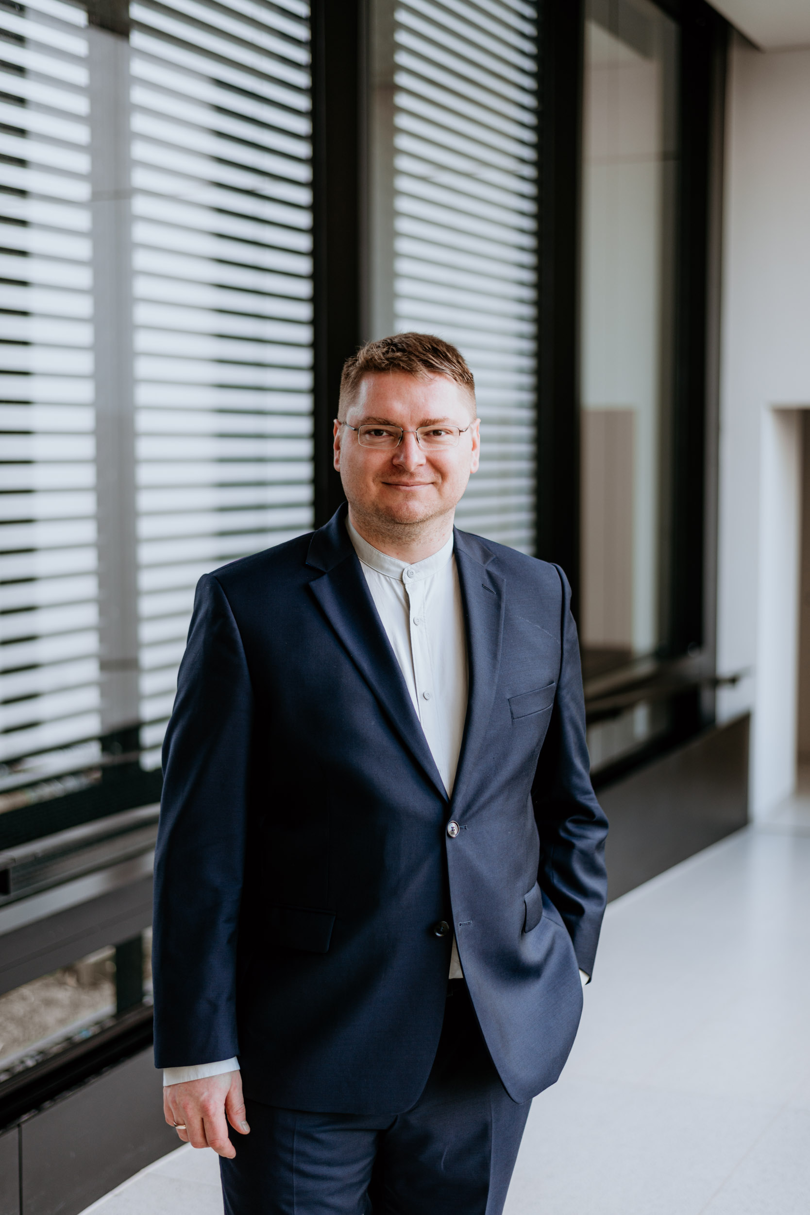
Franz Giessibl
University of Regensburg, Germany Talk: Exploring the sub-picoscale in forces and distances
Abstract:
Abstract: Scanning probe microscopy has brought us experimental access to the world of single atoms. The atomic force microscope (AFM), offspring of the scanning tunneling microscope (STM), has rapidly found wider applications than the STM because it allows to image any sample without requiring electrical conduction. However, the spatial resolution of AFM initially was inferior to the one of the STM. In the last years, the AFM’s resolution has been boosted far beyond STM, e.g. when imaging organic molecules and even revealing electron clouds within single atoms. Height resolution reaches sub-picometer values [1], and Fig. 1 shows force contrasts beyond piconewtons [2,3]. The origin of the forces covers chemical bonding forces, Pauli repulsion forces as well as exchange forces. Is this the end of the development of AFM? The answer is a clear no, as there are some inspiring challenges that require sizable future developmental efforts.
Bio:
Education:
1981-1982: University of Applied Sciences Munich, Microengineering, Prediploma,
1982-1988: Technical University Munich and Federal Institute of Technology Zurich, Physics, Diploma thesis with Prof. Dr. Gerhard Abstreiter in experimental semiconductor physics,
1988-1992: Ludwig-Maximilians-University Munich / IBM Research Group Munich Dr. rer. nat. in experimental physics with Prof. Dr. Gerd Binnig in Atomic Force Microscopy,
2001: Universität Augsburg, Habilitation, Habilitation-thesis "Progress in Atomic Force Microscopy" (2000).
Professional Experience:
1981-1988: Numerous summer jobs with International Business Machines Corporation, Max Planck Institute and others,
1992: Postdoctoral Fellow IBM,
1992-1994: Park Scientific Instruments Inc., Sunnyvale, California: UHV R&D Scientist, Senior Scientist and Director of UHV Products,
1995-1996: McKinsey&Company, Inc.: Senior Associate,
1996-2006: Universität Augsburg, Experimentalphysik VI: Leader scanning probe microscopy group and lecturer,
2005: Offer for a Chair in Experimental Physics at University of Bristol (England), Offer for a Chair in Experimental Physics at University of Regensburg,
2006 - today: Full Professor at Universität Regensburg, Naturwissenschaftliche Fakultät II - Physik, Institut für Experimentelle und Angewandte Physik.
Andrea Konečná
CEITEC BUT, Czech Republic Talk: Insulating and Metallic Nanostructured Vanadium Dioxide Probed by Electron Energy-Loss Spectroscopy
Abstract:
In the talk, I will explain how a scanning transmission electron microscope (STEM) equipped with an electron energy-loss spectrometer (EELS) can be used to analyze structural, chemical, and functional properties of matter with spatial resolution down to single atoms. I will demonstrate the capabilities of STEM-EELS in the study of nanostructures made of vanadium dioxide (VO2), which is a material exhibiting a phase transition accompanied by a lattice transformation and an abrupt change in optical and electronic properties from insulating to metallic. The STEM-EELS experiments with in-situ heating helped us to analyze the phase transition in individual VO2 nanoparticles and reveal its dependence on the nanoparticle size.
Bio:
Andrea Konečná is a junior group leader and lecturer at the Institute of Physical Engineering of the Brno University of Technology. She is dealing with research topics at the interface of nanophotonics and state-of-the-art electron microscopy and spectroscopy. In particular, she is interested in the theoretical description of the interaction between fast electrons and optical excitations in nanostructures, such as plasmons, phonons, and excitons, or in electron beam shaping by optical fields.
Martin Švec
Institute of Physics of the Czech Academy of Sciences, Czech republic Talk: Light, tips and molecules: SPM on the path to direct nano-optical measurements
Abstract:
We use tip-enhancement effect in SPM junctions to pursue hard-to-access interactions between light and matter at submolecular scale and perform visualization of the fascinating inner processes of nanoscopic emitters.
We use electroluminescence, photoluminescence and tip-enhanced Raman spectromicroscopy together with the STM, STS and AFM. This provides an opportunity to probe the eigenmodes, charges, vibronics and temporal evolution of the transient states in individual molecues.
Bio:
Martin Svec is currently a group leader of the Scanning Probe Luminescence Microscopy Lab at the Institute of Physics, Czech Academy of Sciences. His team is performing fundamental research in the field of nanocavity-enhanced spectromicroscopy using cryogenic ultrahigh-vacuum SPM instrumentation with optical access. The results of the research are referenced at https://splm.fzu.cz/publications2.php
Grzegorz Korpala
MiViA GmbH, Germany Talk: AI in Microstructure Characterization: Analyzing Real-World Data
Abstract:
In the metallurgical realm, accurate microstructural characterization, especially the measurement of grain size, is pivotal for understanding material properties. This presentation embarks on a detailed analysis comparing the data quality of microstructure images of metals, predominantly steel and aluminum, produced in our laboratory versus those uploaded by external users. Preliminary assessments showcase discernible differences in clarity, resolution, and accuracy between the two categories. Influencing factors on the quality of these images include the sophistication and calibration of microscopy equipment, proficiency of the operator, lighting conditions, and sample preparation methods. User-uploaded images tend to manifest variations, often stemming from diverse equipment standards, varying sample preparation techniques, and potential lapses in capturing methodologies. The importance of data homogenization in this context cannot be overemphasized. Ensuring uniformity in the data sets is essential for reliable grain size measurements, thereby providing a consistent baseline for metallurgical analysis. Addressing these variances is a cornerstone for advancing our understanding of metal behaviors and properties, enabling both researchers and industries to make informed decisions and predictions based on precise microstructural assessments.
Bio:
Education:
1991–1999: Grundschule Krakau, 1999–2003: Lyzeum No. 7 Krakau (Abitur),
2003–2009: AGH Berg und Hüttenakademie Krakau (Master's thesis),
2007–2009: TU Bergakademie Freiberg (Diploma thesis),
2017: Doctorate (Dr.-Ing.) from TU Bergakademie Freiberg
Professional Experience:
2008 – 2009: ThyssenKrupp Steel AG, Duisburg – Materials Competency Center, Part-time (Diploma thesis),
2009 – 2023: Group Leader and Research Associate, TU Bergakademie Freiberg, Institut für Metallformung,
2023 – Present: Founder and CTO, MiViA GmbH
Areas of Expertise
Material Characterization, Artificial Intelligence in Material Characterization
Henning Markötter
BAM Berlin, Germany Talk: Tomographic Imaging Capabilities with hard X-Rays at BAMline (Bessy II)
Abstract:
The BAMline at the synchrotron X-ray source BESSY II (Berlin, Germany) is supporting researchers, especially in materials science. As a non-destructive characterization method, synchrotron X-ray imaging, especially tomography with hard X-Rays, plays an important role in structural 3D characterization. The imaging capabilities allow for in-situ and operando experiments. In this presentation the equipment, data handling pipeline as well as various examples from material science are presented.
Bio:
Dr. rer. nat. Henning Markötter studied physics at the Technical University Berlin, Germany. In 2010 he joined the “Imaging Group” at HZB and did a PhD until 2013, in which he was working in the field of water transport processes in PEM fuel cells by means of synchrotron and neutron imaging. Now he is at the Federal Institute of Materials Research and Testing as a beamline scientist at the BAMline at the electron storage ring Bessy II responsible for the CT activities.
Pavel Plevka
CEITEC MU, Czech Republic Talk: Enterovirus cell entry and genome delivery
Abstract:
Enteroviruses from the family Picornaviridae cause a wide range of diseases, including the common cold, hand-foot-and-mouth disease, encephalitis, and paralysis, with major health, economic, and societal impacts. To initiate infection enteroviruses must deliver their genomes into the cell cytoplasm. However, the mechanism how enteroviruses cross the endosome membrane into the cytoplasm remains unknown. We discovered that enterovirus-containing endosomes deform, rupture, and release virus particles into the cytoplasm. Moreover, we show that endosome disruption also occurs in uninfected cells and after the endocytosis of very low-density lipoprotein and demonstrate that this process depends on N-WASP-regulated actin polymerization. Our results identify endosome membrane remodeling as a new target for the development of therapeutics against enterovirus infections.
Bio:
Michal Kozubek
FIT MU, Czech Republic Talk: AI in microscopy: Coping with multiple scales and multiple modalities
Abstract:
Artificial intelligence (AI), especially deep learning, has already become an indispensable part of microscopy image processing and analysis. It can help with the improvement of image quality (suppressing noise and blur), synthesis of missing information (software super-resolution or computation of an image as if acquired in a different mode such as bright-field from fluorescence) as well as finding objects in the acquired images (object detection, segmentation or tracking). AI methods for these tasks typically outperform the traditional ones and, moreover, they can cope with multiple scales (in microscopy this corresponds to various magnifications) and multiple modalities (in microscopy this means multiple acquisition modes like bright-field, fluorescence, phase contrast, DIC in light microscopy). The talk will show the capabilities of modern AI methodology on an example of live cell image analysis in time-lapse light microscopy.
Bio:
Michal Kozubek is a full professor at Masaryk University (Brno, Czech Republic). He is a head of the Centre for Biomedical Image Analysis (CBIA), and former Dean of Faculty of Informatics. His research concentrates on biomedical image acquisition and analysis with a special focus on spatiotemporal studies. He tries to interconnect biological imaging, medical imaging and artificial intelligence, especially in oncology related research and personalized patient care. He is a fellow of AI-Czechia, vice-president of Czech-Bioimaging research infrastructure (part of Euro-Bioimaging) and a coordinator of Czech Centre for Artificial Intelligence in Oncology. He is a member of MICCAI, IEEE Signal Processing Society, European Microscopy Society as well as a Board member of the MICCAI special interest group on challenges. He has (co-)authored over 100 peer reviewed full papers in international conferences and journals.
Alexander von Appen
MPI CBG, GermanyTalk: Correlating Structure and Dynamics to Understand Nuclear Self-Assembly.

Dirk van der Wal
TESCAN Group, Czech Republic Talk: Correlative Microscopy: from Data to Information
Abstract:
The purpose of analytical instrumentation is to provide information about properties of materials and/or functional structures, such as e.g., battery cathode particles.
Data acquired with a specific analytical instrument can be inconclusive. In such cases, data from other modalities, each also inconclusive, are combined to yield the information of interest.
Such correlative datasets can be multiscale combining data from imaging modalities at different length-scales, or multimodal combining data from different analytical modalities.
Examples of correlative microscopy applications will be discussed.
Bio:
Dirk van der Wal is Chief Marketing Officer at TESCAN GROUP, a.s., Brno, Czech Republic. He joined Philips Electron Optics in 1994, initially as Application Specialist for SEM and EBSD product solutions. He has had multiple Product Management and Product Marketing positions at FEI, ZEISS and now TESCAN, contributing to the development and marketing of emerging Light and Electron Microscopy technologies and methods, such as ESEM in the 90’s, FIB/SEM in the early 2000’s, Automated Mineralogy, Table-top SEMs, Industrial Microscopes such as Digital Light and Confocal Microscopes, and more recently 4D-STEM.
Miroslav Kolíbal
CEITEC BUT, Czech Republic Talk: Monitoring dynamic reactions inside the scanning electron microscope
Abstract:
Our current understanding of catalytic and growth reactions is based on ex-situ (post-growth) characterisations of growth or reaction products, or on monitoring the processes under modified conditions. These conditions usually involve very high vacuum, because it is required by many of the analytical tools we currently use. Indeed, a knowledge obtained in such studies is far from complete and the processes occuring under real reaction conditions may differ, hindering further technological progress. Hence, a large effort exists to develop methodologies that allow to monitor the reaction processes under conditions very close to the real ones. In this contribution, I will shortly describe our efforts towards this goal, challenges that have to be overcome and some of the successful results, namely the formation of 2D materials inside the electron microscope, and monitoring of the oscillatory waves during catalytic reactions by several analytical techniques, including SEM and SIMS.
Bio:
Dr. M. Kolibal has been involved in various research projects dealing with hybrid top-down/bottom-up lithographic approaches for preparation of nanostructures and electron microscopy. He was a visiting scientist at FEI R&D in Hillsboro, Oregon (USA) and at IBM Zurich Research Laboratory (Switzerland), developing in-situ electron microscopy for observation of CVD processes and high-k dielectric templates for directed III-V nanowire growth, respectively. His current research interests include low-dimensional materials, wide-bandgap semiconductors and gas-phase catalytic reactions. A common link in between these research areas is in-situ and in-operando microscopy, which is the main workhorse in his group.
Radim Chmelík
CEITEC BUT, Czech republic Talk: Correlation of hiQPI and LSCM as a tool for revealing molecular mechanisms in cell biology
Abstract:
Holographic Incoherent-light-source Quantitative Phase Imaging (hiQPI) has been established as a powerful, label-free approach to imaging live cells. Cellular biomass density is measured in widefield with unprecedented accuracy. This is why hiQPI can rapidly measure live cells' growth and motility and calculate cell quantitative dynamic morphometry from the image data. Complementing this highly accurate but unspecific image data with highly specific imaging of molecular species with Laser Scanning Confocal Microscope (LSCM) provides a powerful tool for revealing molecular mechanisms in cell biology. To achieve this, we have upgraded a commercial hiQPI microscope with a specimen holder and additional software enabling correlative imaging using Nikon A1R LSCM.
Bio:
JRadim Chmelik is the head of the Experimental Biophotonics research group at CEITEC BUT. He received his PhD in physical and materials engineering from Brno University of Technology for the theoretical description of focusing by diffractive lenses. As a postdoc, he studied methods of 3D imaging in light microscopy. This led him to build, with Zdenek Harna, the first holographic microscope capable of working with completely incoherent illumination. His group focuses on both the development and application of hiQPI technology. His main field of interest is coherence effects in holographic microscopy used for imaging in turbid media and 3D imaging. In 2016, he received the most prestigious Czech Brains award in the Invention category.

Jakub Lázňovský
Talk: Unlocking Insights: X-ray Micro Computed Tomography as a Correlative Imaging Technique
Abstract:
X-ray micro and nano Computed Tomography stands as a versatile tool providing a non-destructive insight into diverse materials in 3D. The challenge is not only to visualize the internal structure of the samples but also quantitatively describe their properties. Therefore, the prevailing trend is to correlate 3D X-ray CT with complementary 2D imaging techniques, that can provide validation of CT data, its extension, or map its elemental composition. In this talk will be presented how can be X-ray CT integrated with various 2D imaging methods, covering data acquisition, processing, and visualization in different research areas.
Bio:
Jakub Laznovsky, a biomedical engineer presently employed at CEITEC BUT, Czech Republic, specializes in X-ray Micro and Nano Computed Tomography. His primary research focus centres around advancing image-processing techniques to facilitate the quantitative analysis of micro and nano CT data. He possesses experience in international collaborations, contributing to diverse scientific projects that involve the characterization of biological samples, 3D printed materials, and composites. He maintains a close cooperation with the Max Planck Institute in Plön, Germany. He will present recent endeavours related to correlating X-ray micro and nano CT with various 2D imaging techniques.
Alexey Boubnov
Talk: Correlative Characterization – Bridging 2D/3D Imaging with Micro-Structuring
Abstract:
We show our correlative method, combining micro/nano-CT, SEM, TEM and light microscopy with laser micro-structuring and FIB, on the same region of interest. The correlation is guided by reference markers, and a crucial aspect is to accurately track these markers at different scales in three dimensions, which becomes increasingly difficult for very small regions of interest. The goals and challenges of our correlative method are shown using a case study with a heterogeneous (Fe,Mo)Ox catalyst.
Bio:
Alexey Boubnov (PhD 2014) is an experimentalist in the field of materials characterization. He has over 10 years of experience in synchrotron X-ray spectroscopy for catalysis research, including experiment design, setup construction, data acquisition and analysis. His recent activities in correlative characterization include correlative sample holder design, image processing and statistical analysis. His special interest lies in complementing experiments with simulations and theoretical models.
Bohuslav Rezek
Talk: 3D multiphase SEM-AFM correlative microscopy
Abstract:
Research investigating the interface between biological organisms and nanomaterials requires nowadays diverse microscopic and spectroscopic methods to elucidate the interaction mechanisms. We will describe a methodology correlating data from atomic force microscope inside scanning electron microscope (AFM-in-SEM). This approach will be demonstrated on multiphase samples of bacteria-nanodiamond-nanometal complexes that are relevant in current life science research. We will discuss principles how a 3D insight into such multiphase samples can be provided by a suitable in-situ cross-correlation of secondary electrons and back-scattered electrons at various energies of primary beam and atomic force microscopy regimes. The obtained knowledge about the microscopic bio-nano-material interactions would hardly be achievable by separate analyses.
Bio:
Bohuslav Rezek graduated from Physics at the Faculty of Mathematics and Physics at the Charles University in Prague in 1996. In 2001 he obtained PhD at the Czech Academy of Sciences (CAS) on the study of charge transport in silicon thin films for photovoltaics by using scanning probe techniques. After PhD he was doing research on diamond devices and biointerfaces in Germany, Switzerland and Japan. In 2006 he became research team leader and Purkyně Fellow at the Institute of Physics CAS. In 2015 he became a head of Physics dept. at the Faculty of Electrical Engineering of the Czech Technical University in Prague. In 2019 he became full professor there. His research is focused on correlative microscopic and micro-spectroscopic analyses of semiconductor and organic materials towards opto-electronic and bio-electronic applications.
Luís Pereira
CENIMAT, Portugal Talk: Cellulose nanocrystals: from self-assembly to electronic, photonic and energy harvesting devices inspired by nature
Abstract:
The presentation will focus on applying cellulose nanocrystals (CNCs) in new electro-optic devices that combine these CNCs with inorganic semiconductors. It will be shown how the control of evaporation-induced self-assembly (EISA) of CNCs in aqueous suspensions can enable unique properties when using CNCs in field effect transistors and photodetectors sensitive to the polarization of light towards nature-inspired hybrid electronics and photonics. Interaction with different ions affects the electrochemical properties of CNCs films and their self-assembly behavior compared to pristine ones. Understanding the fascinating properties of this material and how soft matter can be combined with solid-state physics requires different characterization techniques that may be correlated and used to create models predicting the optical response of such hybrid devices.
Bio:
Luis Pereira is a PhD in Microelectronics and Optoelectronics and Associate Professor at NOVA University Lisbon. Current research interests are in the design and synthesis of 1D, 2D and 3D inorganic and hybrid nanostructures, cellulose nanocomposites, functional micro and nanofibers, and their integration of electronic, chromogenic, and energy-related devices. He is the Technical and Scientific Director of the collaborative laboratory AlmaScience, Coordinator of the Materials Engineering College-Portuguese Professional Engineer Association – South Region, Vice President of the Portuguese Materials Society (SPM), and was recently elected as a member of the executive committee of FEMS.
Adrian Nievergelt
CBI Max Planck Institute, Germany Talk: The eukaryotic proteome as a driver for multi-correlative method development
Abstract:
Eukaryotic proteins constitute some of the most complex molecules on earth. They depend not only on their primary peptide sequence for their function, but frequently require a stunning array of post-translational modifications, oligomerization, ultrastructure, compartmentalization and dynamic behaviour to fulfil their function. Using examples from the cilio-basal apparatus, I will show how we can observe these different properties of biomolecules by high speed atomic force microscopy, fluorescence microscopy, mass spectro- and photometry as well as cryo-electron microscopy.
Bio:
Adrian is an engineer in the broadest sense, interested in all disciplines of engineering. He studied nanosystems engineering at the Swiss Federal Institute of Technology in Zurich, followed by a doctorate at École Polytechnique Fédérale de Lausanne. For his doctoral project, he designed and built high-speed atomic force microscopes, enabling the real-time observation of protein dynamics at molecular resolution. He then joined the Max Planck Institute of Molecular Cell Biology and Genetics, where he is working on the genetics, structural biology, and metabolism of Chlamydomonas.
Satellite event
The 12th European School for Young Materials Scientists aims to bring together young scientists, mainly PhD students, working in the exciting fields of Materials Science, Nanotechnology, Nanoscale Materials, Nanoanalysis, Multi-scale materials characterization, as well as Microscopy.
The first part of School will be a two days event and will count with an inspiring keynote lecture from a prominent scientist in the above-mentioned fields, as well as and most importantly, the very active participation of the PhD students. The event will create a live forum that brings together presentations of the PhD works (oral and posters), intensive discussions, and many opportunities for interaction and networking.
The second part of the School will be organized in close cooperation with the 3rd Correlative Materials Characterization workshop at November 9-10 and all young scientists will be invited to attend the CMC. The company tours at Tescan and ThermoFisher will be organized together with CMC at November 9 in the morning.
ESYMS'23 School PosterRegistration and Abstract Submission:
Registration and Abstract Submission until September 15th, 2023
Abstract template:
Download here
Sponsors
- City of Brno
- South Moravian Region
- TESCAN GROUP, a. s.
- Thermo Fisher Scientific
- Delong Instruments
- deepXscan
- Měřící Technika Morava - Bruker
- NenoVision
- Telight
- ZEISS
[logoshowcase center_mode="true" slides_column="3" cad_id="67"]
Commitee
- Mr. Tomáš Šikola, Brno University of Technology
- Mr. Pavel Tomančák, CEITEC
- Mr. Ehrenfried Zchech, deepXscan
- Mr. Jan Neuman, NenoVision
- Mrs. Lucie Mezníková, Czech Optical Cluster
- Mrs. Pavla Skácelová, Czech Optical Cluster
- Mr. Petr Přikryl, Czech Optical Cluster
- Mr. Jan Neuman, NenoVision
Practical information
You can comfortably get here by air, bus, train or car.
By Air
- Brno – Tuřany Airport: Visitors of Brno can use International Airport Brno-Tuřany, which provides flights to London/Stansted and other charter destinations. Airport is located 12km/20 minutes from the city center and you can easily take a taxi or the express bus, which is directly located outside the entrance to the airport terminal.
- Vienna International Airport: There is no direct flight from Vienna to Brno because of too short distance. Easiest transportation from Vienna airport to Brno is with bus operator RegioJet. Tickets are available to purchase online at the website.
- Václav Havel Airport Prague: Easiest transportation from Prague is with bus operator RegioJet. Tickets are available to purchase online at the website.
RegioJet is a reliable bus and train company. You can also use Ceske drahy (Czech railways). Universal bus/train search engine and public transport planner – www.idos.cz.
By car
Brno is located on the main Czech highway D1 and D2. For highways and some bigger roads it is necessary to have a highway sticker, which is for sale at the border crossing, petrol stations, post offices and in the motor clubs.
Venue

- situated almost in the centre of the city of Brno, in a park on the hill of Kraví hora.
- By public transport you get there by a tram no. 4, tram terminal “náměstí Míru/Masarykova čtvrť”.
- Parking spaces (also for buses) can be found in the square náměstí Míru or at the swimming pool on the hill of Kraví hora.